My First Time Doing Dissections
I have to start this post by saying that if you don't want to see the insides of a sea urchin, you should skip this one and read one of our other blog entries.

For those of you still with me, let me start from the beginning. I had never done any animal dissections in my academic career before Antarctica. Maybe a fetal pig in my high school biology class, but nothing at this level. So when I found out I would be helping with the dissections this season, I got excited. My project looks at microbes, which are so tiny you can't see them with the naked eye, so working with animals big enough to see and learn about up close was thrilling.

In the weeks that we've been here, I've had the pleasure of doing dissections of Odontaster validus (sea star), Sterechinus neumayeri (sea urchin), and even Parborlasia corrugatus (ribbon worm) for one of the lab's projects. These dissections need to happen so that our lab can study these animals in more depth and understand what they are eating in places where methane is present.
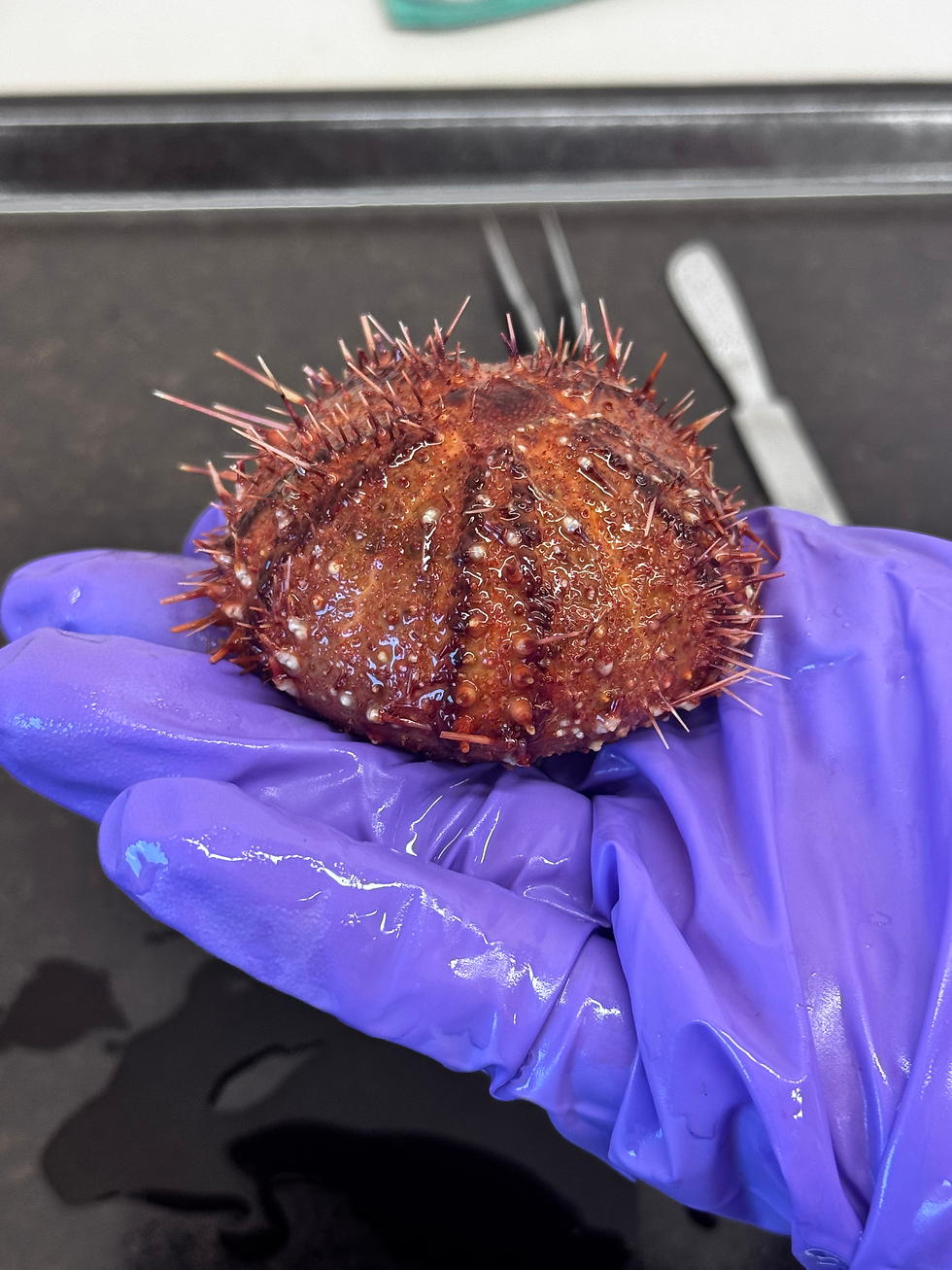

I was struck by how beautiful these animals are, inside and out. Following Paola's guide, I opened the first sea urchin. I didn't expect them to be so symmetrical inside, or to be able to see so much. I could see some eggs, Aristotle's lantern right in the middle (their jaw apparatus), the stomach, and the intestines winding around the inside. It was fascinating to see how everything fit together like puzzle pieces. I was also amazed by how well they hold on to things. A couple of them were even clinging to some of the sea stars collected that day. Sea urchins use their spines for protection and movement, and their tube feet for gripping, anchoring themselves, and gathering food.
This experience really showed me that we are always learning, and that's okay. It also reminded me to be more open to new experiences inside and outside of science, because you never know what might surprise you.
We greatly appreciate and respect all of these animals and their sacrifices. This project will help us understand these communities better and be better prepared to protect them in the future. So to all the sea stars, sea urchins, and ribbon worms of this and past seasons: thank you for your service.


Comments